Photo: disorders.com
Pigs that are NOT healthy/genetic abnormalities
There are many different traits in pigs that should NOT be purposefully chosen as a fixed or desired trait. Many of these pigs who suffer from these genetic anomalies suffer horribly or pass away much earlier than expected because of their genetics. If you are looking at pigs, find out some history (if the person knows any), if there were other pigs, ask how they're doing (assuming this person keeps in touch with past pig parents)....be sure the parents of the pig you are getting are healthy. Many of these traits aren't easily recognized unless you know what you are looking for, this is why we created this particular page. The examples below are scientifically proven to be mutated genes, traits that can be problematic. The defects listed aren't a complete list, we will continue to build this page as we are able to find credible information to include. At the bottom of the page is a PDF file with various studies that discuss how some of these abnormalities came to be considered as genetic defects.
Mulefoot pigs
Mulefoot pigs is a desired "unique" pig. What people don't know is that the mule foot is also a genetically defective pig. Watch this video about a ONE year old pig with mule foot.
Dwarfism
People who are seeking a "dwarf" pig may initially be excited they have a pig that MAY stay small....what they don't know is that these pigs rarely live past age 2. Dwarfism is a genetic mutation and isn't something that anyone should breed towards. Pigs that have litters that have a dwarf should NOT be breed because these pigs will NOT live long. What does that leave? A heartbroken family.
The scientific community have identified a naturally occurring, dominant mutation that causes dwarfism in domestic pigs (Sus scrofa). With a positional candidate gene approach, the dwarf phenotype was shown to be a result of a single amino acid change, G590R, in the alpha1 (X) chain of type X collagen. Type X collagen is a homotrimer of alpha1(X) chains encoded by the COL10A1 gene, which is expressed in hypertrophic chondrocytes during the process of endochondral ossification. An amino acid substitution at the equivalent position in human type X collagen, G595E, has previously been shown to cause Schmid metaphyseal chondrodysplasia (SMCD), which is a relatively mild skeletal disorder associated with dwarfism and growth plate abnormality. Consistent with the clinical phenotype of SMCD patients, radiological and histological examination of the dwarf pigs revealed metaphyseal chondrodysplasia in the long bones. Yeast-based, two-hybrid protein interaction studies and in vitro assembly experiments demonstrated that the amino acid substitution interfered with the ability of the mutated collagen molecules to engage in trimerization. This work establishes that the chondrodysplastic dwarf pigs by genetic, biochemical, radiological and histological criteria provide a valid animal model of SMCD.
Known genetic defects that are linked to dwarfism in pigs:
http://www.gse-journal.org/articles/dwarfism-effects-pigs
A study about this genetic defect: http://minipigs.dk/uploads/media/Rethink_-_Article_6_02.pdf
| genetic_dwarfism.pdf |
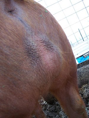
Cryptorchidic boar
Photo credit: sugarmtnfarm.com
Cryptorchidism
There is a condition called Cryptorchidism in males with "retained" or undescended testicles. This may be one or both testicles and I have heard of unsuspecting pig parents being fooled by someone saying a pig is neutered when in fact, the pig is far from neutered. The procedure to locate and remove the testicles in animals with this condition is much more difficult than the standard neutering procedure. The veterinarian needs to be skilled and proficient in mini/potbellied pigs to reduce the risks of a native or disastrous outcome. An experienced vet would be ideal, but this isn't always possible. Having your vet contact an experienced veterinarian like Dr. John Carr of the UK or Dr. Wilbers of Quarkertown Vet Clinic in Pennsylvania would be a great option if your vet wanted to consult a more experienced veterinarian beforehand. Most university vet hospitals have experienced vets on staff as well. This is a genetic disorder, it can be passed on to offspring and can be a result of inbreeding too. Breeders who pass these pigs off as neutered should be informed that these pigs are NOT neutered and if they have proof of surgery, that veterinarian who claimed to do a procedure to remove the testicles, should be reported to the veterinarian licensing board. This is unacceptable and people who are passing pigs on as being neutered knowing they're not ought to be ashamed of themselves and these dishonest people also need to be reported to someone. Even if you let others know of their scam, that may prevent others from being fooled. Since this is a genetic disorder, the cycle can continue for quite some time and lead to major health complications in pigs who are identified as neutered when they're not.
Aggression is a big concern in intact pigs and boars cannot be trusted. So these morally constipated people who are flat out lying to people can literally put others lives in danger. An intact boar can fatally wound a person given the right circumstances and opportunities. It still isn't the pigs fault, as their hormones drive their behavior. If you were told your pig was neutered and see scrotal sacs on the hind end? Your pig is likely NOT neutered. Perhaps he had one or both testicles retained and they finally dropped or they weren't very prominent to begin with. If your pig is displaying classic boarish behavior like humping any/everything, you may want to have your vet check your pig out to be sure your pig is not intact. There is a difference between a pig with behavioral issues and a pig with hormonal issues. A story about a boar that "attacked" both husband and wife can be read by clicking here. (This story is very questionable to me and this pig did NOT need to be euthanized, a neuter would have likely solved the problem)
Diagnosing an animal as bilaterally cryptorchid, as opposed to having been previously desexed, is generally difficult to do on the basis of testicular palpation alone. After all, in each case, there will be no testicles felt in the scrotal sac. The only time that testicular palpation may be able to assist in the diagnosis of bilateral cryptorchidism is if one or both of the retained testicles is outside of the abdomen and located within the inguinal canal or prescrotal area and, therefore, able to be palpated by the vet.
Palpation of the empty scrotum may give the veterinarian clues about whether or not a pig was desexed (castrated) as opposed to being bilaterally cryptorchid. Desexed animals tend to have a thick ball or nub of scar tissue within each of the scrotal pouches. But this is not a foolproof method to be 100% certain a pig has been neutered.
This condition, left untreated, can actually lead to complications if not treated. Pigs with cryptorchidism are at a much higher risk for developing testicular tumors as well as these conditions.
As mentioned above, animals with bilateral cryptorchidism might be infertile and incapable of making viable sperm, however, their testicles are still capable of producing high levels of the masculinising hormone: testosterone. Desexed males, on the other hand, have very little testosterone. Bilaterally cryptorchid animals with plenty of testosterone are therefore much more likely to develop the kinds of "male" testosterone-dependent body features normally attributed to an entire animal.
It is sometimes possible for your veterinarian to locate bilaterally undescended testicles using ultrasound technology. Many clinics have access to diagnostic ultrasound these days and a skilled operator may be able to locate the undescended testicles. Note, however, that undescended testicles are often small and can be very difficult to find and that a negative result on ultrasound does not mean that the animal is not cryptorchid.
Sometimes a diagnosis of bilateral cryptorchidism (as opposed to monorchism, anorchism or prior castration) can only be made by surgical exploration. In these situations, the animal is placed under a general anaesthetic and its entire abdomen, groin and scrotal region is shaved and surgically prepared. Because the missing testicles might be very small (retained testicles are often a lot smaller than scrotal testicles are) and could be located anywhere from the abdominal cavity, behind the kidney, through the inguinal canals to the prescrotal regions, the vet may have to make several incisions to find them. The vet might make an incision into the abdominal cavity, only to discover that one or both of the retained testicles' spermatic cords (vas deferens and testicular blood vessels) disappears into the respective inguinal canal. The vet will then need to make a new incision into the appropriate right and/or left inguinal (groin) region/s to locate the missing testicle/s. This is the more complicated surgery mentioned above.
Excessive estrogen production, as can occur in male animals with certain testicular tumours or female animals with estrogen-secreting ovarian tumours or estrogen-secretory ovarian follicular cysts, can cause the feminising syndrome. In its severe form, it can also produce signs of estrogen toxicity, resulting in severe, often life-threatening effects on the animal's bone marrow.
Aggression is a big concern in intact pigs and boars cannot be trusted. So these morally constipated people who are flat out lying to people can literally put others lives in danger. An intact boar can fatally wound a person given the right circumstances and opportunities. It still isn't the pigs fault, as their hormones drive their behavior. If you were told your pig was neutered and see scrotal sacs on the hind end? Your pig is likely NOT neutered. Perhaps he had one or both testicles retained and they finally dropped or they weren't very prominent to begin with. If your pig is displaying classic boarish behavior like humping any/everything, you may want to have your vet check your pig out to be sure your pig is not intact. There is a difference between a pig with behavioral issues and a pig with hormonal issues. A story about a boar that "attacked" both husband and wife can be read by clicking here. (This story is very questionable to me and this pig did NOT need to be euthanized, a neuter would have likely solved the problem)
Diagnosing an animal as bilaterally cryptorchid, as opposed to having been previously desexed, is generally difficult to do on the basis of testicular palpation alone. After all, in each case, there will be no testicles felt in the scrotal sac. The only time that testicular palpation may be able to assist in the diagnosis of bilateral cryptorchidism is if one or both of the retained testicles is outside of the abdomen and located within the inguinal canal or prescrotal area and, therefore, able to be palpated by the vet.
Palpation of the empty scrotum may give the veterinarian clues about whether or not a pig was desexed (castrated) as opposed to being bilaterally cryptorchid. Desexed animals tend to have a thick ball or nub of scar tissue within each of the scrotal pouches. But this is not a foolproof method to be 100% certain a pig has been neutered.
This condition, left untreated, can actually lead to complications if not treated. Pigs with cryptorchidism are at a much higher risk for developing testicular tumors as well as these conditions.
- Testicular torsion (twisted testicle)
- Testicular cancer (testicle cancer)
- Male feminizing syndrome
- Bone marrow hypoplasia and pancytopenia- estrogen toxicity
- Excessive testosterone production- this can lead to super aggressive behaviors.
As mentioned above, animals with bilateral cryptorchidism might be infertile and incapable of making viable sperm, however, their testicles are still capable of producing high levels of the masculinising hormone: testosterone. Desexed males, on the other hand, have very little testosterone. Bilaterally cryptorchid animals with plenty of testosterone are therefore much more likely to develop the kinds of "male" testosterone-dependent body features normally attributed to an entire animal.
It is sometimes possible for your veterinarian to locate bilaterally undescended testicles using ultrasound technology. Many clinics have access to diagnostic ultrasound these days and a skilled operator may be able to locate the undescended testicles. Note, however, that undescended testicles are often small and can be very difficult to find and that a negative result on ultrasound does not mean that the animal is not cryptorchid.
Sometimes a diagnosis of bilateral cryptorchidism (as opposed to monorchism, anorchism or prior castration) can only be made by surgical exploration. In these situations, the animal is placed under a general anaesthetic and its entire abdomen, groin and scrotal region is shaved and surgically prepared. Because the missing testicles might be very small (retained testicles are often a lot smaller than scrotal testicles are) and could be located anywhere from the abdominal cavity, behind the kidney, through the inguinal canals to the prescrotal regions, the vet may have to make several incisions to find them. The vet might make an incision into the abdominal cavity, only to discover that one or both of the retained testicles' spermatic cords (vas deferens and testicular blood vessels) disappears into the respective inguinal canal. The vet will then need to make a new incision into the appropriate right and/or left inguinal (groin) region/s to locate the missing testicle/s. This is the more complicated surgery mentioned above.
Excessive estrogen production, as can occur in male animals with certain testicular tumours or female animals with estrogen-secreting ovarian tumours or estrogen-secretory ovarian follicular cysts, can cause the feminising syndrome. In its severe form, it can also produce signs of estrogen toxicity, resulting in severe, often life-threatening effects on the animal's bone marrow.
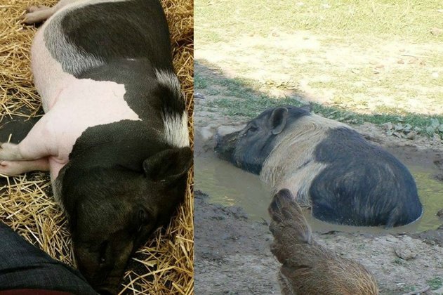
Hair/Skin
The basis of hairlessness has been established hereditary (hypotrichosis) by Roberts and Carol (1931). This condition which is to be distinguished from a similar one due to a deficiency of iodine , is due to a single autosomal recessive gene, which reduces the number of hair follicles.
Another type of hypotrichosis has been described by Meyer and Drommer. In their case an autosomal dominant gene is involved. This character is lethal, as homozygous hypotrichotic piglets die within 10 days. The vitality of heterozygous individuals is also reduced.
Disturbances in the arrangement of the hair, known as whorls or "roses", occur mainly along the spinal column. They have been explained by the complementary action of two dominant genes.
Several kinds of skin defects, of a hereditary nature, have been reported in the pig. A condition known as epitheliogenesis imperfecta is characterized by areas of missing epidermis of variable and irregular size. The condition is semi-lethal, as affected piglets usually die within three days but may survive if the abnormal area is small and the animal properly handled. The first case was reported by Nordby (1929) who considered the abnormality to be probably genetic. This has been confirmed by segregation results obtained by Sailer (1955), which correspond to a single autosomal recessive gene.
The occurrence of melanotic skin tumours was first studied by Nordby (1933), who came to the conclusion that the defect is inherited, but with an unclear mode of inheritance. This has later been confirmed by HKoo et al. (1979) who were able to increase the frequency of the defect by selection in a line of miniature pigs. Their data suggest that the hereditary basis is polygenic and similar to that reported in humans.
A hereditary basis of the transient skin disease known as pityriasis rosea has for the first time been suggested by Wellman (1953) who was able to exclude infectious agents as possible causes. The disease begins with the apparition of a few hyperhaemic patches on the underside of the animal. These rapidly spread in a circular fashion and then join together to form large circular marks similar to those found in ringworm. The disease in the first weeks of life and generally lasts until 3-4 months of age. The condition is thought to be widespread of a hereditary origin and may go unnoticed by the breeder as general health is not impaired.
Skin lesions of a different origin may also appear in the first or second week of life. This disease, known as dermatosis vegetans, differs from the preceding one in that it also affects the feet and the lungs. The pig usually die within four to six weeks from either pneumonia or secondary bacterial infections. Fatla et al. (1961) showed dermatosis vegetans to be a semi-lethal hereditary disease due to a single autosomal recessive gene, a hypothesis which also fits the Gobservations of Done et al (1967). The disease has also been reported in Austria by Gawisching et al. (1974) who tried unsuccessfully to obtain affected animals from matings between parents known to have given defective progeny. This genetic defect seems to affect mesodermal tissue selectively (dermis, intestinal, lymphoid tissue, Jtonsils and pulmonary lymph nodes) as shown by histological observations by Jericho (1974). The club-foot syndrome, reported by Larsson (1953) and later shown to be inherited as a autosomal syndrome, reported by A (1953) single recessive gene, is likely to be the disease later described as dermatosis vegetans.
Head/Skull
A common skull defect in pigs is brain hernia, which is due to a cleft in the skull, through which meninges may protrude (meningocele) or meninges and brain tissue (encephalocele).
Hydrocephalus is an enlarged head condition which results from an excess of cerebrospinal fluid either in the brain ventricles (internal hydrocephalus) or in the cranial cavities (external hydrocephalus). The defect is variable in expression and has been found associated with tail and sometimes W hair and rudimentary light-coloured skin, in the Duroc breed, by Blunn and Hughes (1938) who showed it to be due to a single autosomal recessive gene.
According to the review by Koch et al.(1957), the exact mode of inheritance for cleft lip, jaw and palate (cheilognathopalatoschisis) is uncertain. A simple recessive gene has been suggested by Norodd (1958) and a recessive gene with incomplete penetrance by Labik (1972). Non-genetic factors may also be involved as shown by the breeding experiment of Butz and Meyer (1960).
Complete absence of the lower jaw (agnathia) has been reported by Keller (1941), but the inheritance of this lethal condition has not been investigated.
A lethal factor, supposed to be recessive, is held responsible for the occurrence of bilobed ears, an abnormality which is sometimes accompanied by cleft palate and hind leg malformations (Annette 1938).
Limbs
Absence of one, two or four legs has been reported in pigs. The absence of the four is a lethal condition, which has been described by Johnson and Lush (1939) and shown to be due to a single autosomal recessive gene. The absence of one or two legs has several times been described in grown pigs. From the work reviewed by Koch et al. (1957) it can safely be concluded that the three-legged condition is due to a single autosomal recessive gene, whereas the less common two-legged condition (apodia) is genetically unclear.
The absence of toes (adacytlia) has also been described in pigs. The absence toes has also been described in either alone or
associated with several other abnormalities (Butz & Schnelle 1951 ; BEER, 1962). A hereditary basis is probable but has not yet been clearly established.
A hereditary defect leading to unequal toes has been described by Nordby (1939). Its exact hereditary basis, however, remains to be explained.
A short-leg syndrome has been shown to be inherited as a single recessive gene by Swiger (1981).
Atticus's Story
This is Atticus. Mo Money For Pigs was contacted to help this family with unexpected vet bills for this guy. "After a radiograph and a CT we are left we some bad news. His leg is broken in two places as a result of IOHC (incomplete ossification of the humeral condyle). the cause? Malnutrition. Genetics." ~MMFP 11/2016
Unfortunately as Atticus's leg was healing, another leg broke and the decision was made to end his suffering. A lifetime full of broken legs is not a good quality of life. His mama, Jodi, went above and beyond to make him comfortable for the short time he was here on earth. Rest in paradise sweet angel.
It is unknown if malnutrition or genetics caused this, but either one could be the reason why his bones were weak. Nonetheless, it is a condition that can be caused by breeding pigs with poor genetics.
Multiple Abnormalities
Various skeletal anomalies may oDccur on the same animal. Such is the case of the Pulawska lethal factor described by A single
Dabczewski (1949) and inherited as a auto-somal recessive gene. Various malformations affect the cranium bones, the vertebral column (vertebral fusion) and the ribs, and several internal organs (liver, pancreas, kidney and intestine are larger than normal, and lungs are rudimentary).
Lameness (or the so-called leg weakness syndrome) occurs frequently in the modern pig. Various forms of skeletal lesions (osteochondrosis, epiphysiolysis, arthritis) are usually at the origin of this defect. An extensive literature is devoted to the subject, and many authors have suggested that the disease is partly of hereditary origin. A moderate heritability is usually found for leg weakness, when it is assessed either visually or radiologically.
Eyes
Lack of pigmentation of the iris leads to the "glass-eye" defect also called heterochromia iridis. An account on the heredity of this condition has been given Gelati et al (1973). In the first work, Durr (1937) concluded to an incompletely dominant gene, as only about 50 percent of the heterozygous individuals Gelati et al. conclude that, in their herd of miniature pigs, the defect is due to an autosomal recessive gene (het), whose expression is variable and in particular is influenced by the white color gene. The two hypotheses can be reconciled if one assumes, as suggested by Gelati, that pigs with bilateral heterochromia are homozygous at the Het locus and unilateral or partial heterochromia, vary frequently in Durr's data, may appear in heterozygous individuals.The gene may thus be considered as truly recessive for bilateral heterochromia.
Congenital blindness is of frequent occurrence in pigs and generally the result of various degrees of microphtalmia. Non-genetic causes may be implied in eye defects, especially lack of vitamin A. Several workers however have shown that a genetic basis exists for blindness in pigs. The various cases studied have been reviewed by Koch et al. (1957) who observe that their own investigations, on the progeny of a blind boar, do not always confirm previous ones and conclude that further research is needed in order to clarify the heredity of the condition.
Cyclopia is an abnormality found in several species and shown to be hereditary in guinea pig and rabbit. In the pig, cyclopia often goes along with several other abnormalities and its heredity remains to be clarified. (Koch et al 1957)
Neurological and neuromuscular disorders
Congenital tremor (or myoclonia) is a frequent disease affecting the central nervous system of pigs, and it may have several different causes. In the taxonomy proposed by DONE (1976 a), those forms of tremor in which morphological lesions are found are called type A. Two of these are of genetic origin, namely AIII, which is due to a sex-linked recessive and AIV, which is due to an autosomal recessive gene.
A congenital motor defect, demonstrated by clinical signs of ataxia and perverse movements, with no morphological defect in the central nervous system at birth, but dysplasia of the cerebellar cortex in older pigs, is inherited as a single autosomal recessive trait in large white pigs and recently confirmed in Yorkshire pigs. (Rimaila-Parnanen 1982)
Splayleg is a condition of newborn piglets in which the hind legs and sometimes the forelegs tend to splay sideSwards and fonvards as a result of muscular weakness. In their reviews of the subject splayleg has a genetic basis, probably polygenic, though sometimes it occurs only in males and may be due to a sex-linked gene with variable penetrance (Lax 1971) Myofibrillar hypoplasia has sometimes been observed in splayleg (though it may also be found in’ normal pigs) and indicates a retardation in the development of the leg muscles as a consequence of a possibly general neuromuscular dysmaturity.
PSE and sudden death are also connected with a genetic defect known as malignant hyperthermia (MH), a syndrome found in several other species including Man and which is triggered by halothane anesthesia. A rapid rise in body temperature, muscular rigidity and blotchy cyanosis of the skin are the most obvious symptoms. Death normally ensues if anesthesia is continued. Click here to read more about PSS (porcine stress syndrome).
Internal organs
Atresia ani (closure of the anal outlet) is one of the most frequent abnormalities encountered in pigs. Either one recessive gene with or two dominant have been as the incomplete penetrance or 2 incompletely dominant genes have been proposed as the most likely genetic explanations. Neeteson (1964), from observations on 36 litters, ruled out six different genetic explanations and retained as a provisional conclusion a two dominant genes hypothesis. The frequency of the defect in affected litters observed by Triebler et al.(1964) agrees with the hypothesis of a recessive with 50 p.penetrance. The linkage mentioned between atresia ani and thickleg by Walther al. (1932) awaits further confirmation. Many inbred pigs have suffered from this condition, some don't survive more than a couple of days. Females who lack an anus will usually rupture through a wall and naturally create a way for feces to be excreted. Males, on the other hand, will sometimes do the same, but male pigs tend to have more complicated issues since they don't have a thin membrane into a vaginal cavity like the girl pigs do, and typically because of the inability to defecate, they pass away within a few days. There have been pigs who have undergone successful procedures to "create a butt-hole" or realign the existing tract and create an opening for the feces to be able to be excreted.
A different abnormality with similar consequences, aplasia of the anal sphincter, Hamori (1965) who considers it as a semilethal defect has been reported by A hereditary whose transmission still remains to be clarified.
Scrotal (or inguinal) hernia, a protrusion of the intestine into the scrotum (or through the inguinal canal), is probably the most frequent hereditary abnormality in the pig. Scrotal hernia is a sex-limited defect, whereas inguinal hernia is found in both sexes. Inguinal hernias in females has been reported to occurring conjunction with abnormal ovaries.
Umbilical hernia is a less frequent type of hernia; its frequency is higher in females than in males. Koch et al.(1957) mention studies showing the condition to be hereditary, possibly of a dominant type.
To read a copy of this series of studies, you can download the PDF file below.
| genetics_review-pigs.pdf |