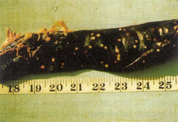
Melioidosis. Multiple abscesses in the spleen of a pig.
Melioidosis
Melioidosis is an infectious disease of pigs, goats and occasionally of other animals caused by Pseudomonas pseudomallei, present in several tropical and subtropical countries in the Asia-Pacific, Middle East and Caribbean regions. It causes fatal infections in a significant proportion of infected humans, especially those who are immuno-compromised or have intercurrent disease.
Transmission : Infection occurs by ingestion of the infective material containing Pseudomonas pseudomallei, contamination of wounds or abrasions of the skin or perhaps bites of insects. Important sources of infection are rodents, contaminated swamps or muddy water. The organism can live up to three months in shaded soil.
Antemortem findings :
Postmortem findings :
Judgement : Carcasses suspected to be infected with melioidosis should be isolated and retained, and the affected tissues, preferably an excised unopened lymph node and the spleen with abscesses should be sent to the appropriate laboratory for examination. Diagnosis is made on the basis of isolation, cultural examination and pathogenicity test. If there are no facilities for retention in isolation, the carcass suspected or tentatively diagnosed to have melioidosis should be condemned and properly disposed of. Persons handling the suspect carcass or material should disinfect their hands, forearms, knives and other contaminated equipment. If the laboratory report is negative the carcass has to be treated on the basis of the lesions present and the subsequent diagnosis.
Differential diagnosis : Tuberculosis, non-specific purulent conditions. Caseous lymphadenitis in sheep and goats and actinobacillosis in sheep.
Source: http://www.fao.org
Transmission : Infection occurs by ingestion of the infective material containing Pseudomonas pseudomallei, contamination of wounds or abrasions of the skin or perhaps bites of insects. Important sources of infection are rodents, contaminated swamps or muddy water. The organism can live up to three months in shaded soil.
Antemortem findings :
- Incubation period is variable, longer in pigs than in other animals.
- Fever up to 41°C especially in sheep and goats
- Enlarged lymph nodes, particularly the sub-mandibular nodes in pigs.
- Loss of appetite
- Posterior paresis and evidence of nervous signs
- Abortion and still births
- Orchitis in boars
- Most cases in pigs are chronic and these may not show clinical signs.
Postmortem findings :
- Multiple abscesses in most organs especially in the regional lymph nodes, spleen (Fig. 136) and liver
- The abscesses contain a thick, caseous greenish yellow or off white turgid pus. There is usually no calcification.
- Pneumonic changes in the lungs
- Suppurative polyarthritis, the joint capsules contain fluid and large masses of greenish yellow pus.
- Meningoencephalitis
Judgement : Carcasses suspected to be infected with melioidosis should be isolated and retained, and the affected tissues, preferably an excised unopened lymph node and the spleen with abscesses should be sent to the appropriate laboratory for examination. Diagnosis is made on the basis of isolation, cultural examination and pathogenicity test. If there are no facilities for retention in isolation, the carcass suspected or tentatively diagnosed to have melioidosis should be condemned and properly disposed of. Persons handling the suspect carcass or material should disinfect their hands, forearms, knives and other contaminated equipment. If the laboratory report is negative the carcass has to be treated on the basis of the lesions present and the subsequent diagnosis.
Differential diagnosis : Tuberculosis, non-specific purulent conditions. Caseous lymphadenitis in sheep and goats and actinobacillosis in sheep.
Source: http://www.fao.org